Microscopy
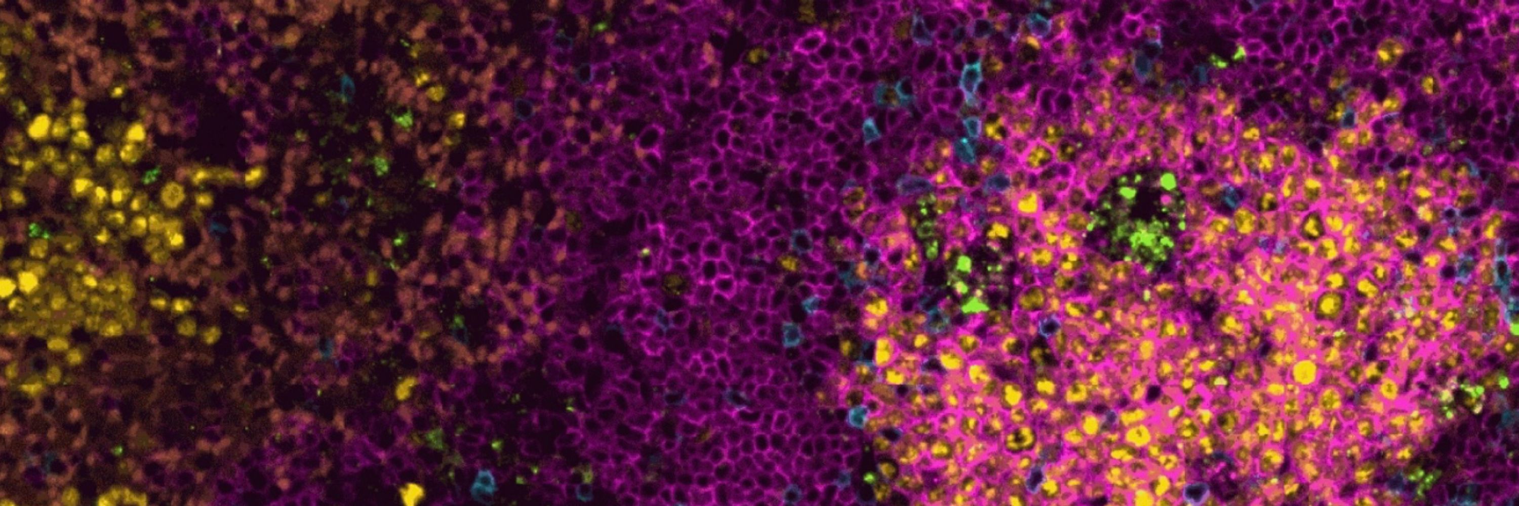
Images brought to you by the La Jolla Institute for Immunology Microscopy and Histology Core
@rarecyte.bsky.social
A one-stop resource for Spatial Biology and Liquid Biopsy solutions. Learn more: https://rarecyte.com/
@lji-lpa.bsky.social
We represent graduate students and postdoctoral scholars at La Jolla Institute for Immunology. We share a passion for immunological research while having fun! 🔬🦠✨🧪🧬🧫
@nat-prunet.bsky.social
Microscopic farmer 🔬🌺 Microscopy core director @UNC Chapel Hill Former plant developmental biologist
@reikawatanabe.bsky.social
Biologist fascinated by and specialized in cellular cryo-electron tomography @ljiresearch.bsky.social. #teamtomo, #cryoET, #virology
@tom-riffel.bsky.social
Group Leader, Center for Inflammation and Autoimmunity Director, Immunometabolism Core La Jolla Institute for Immunology
@geobellward.bsky.social
Light microscopy facility staff with interest in learning and sharing information about sample preparation, image acquisition, and image display best practices. Keen interest in Expansion Microscopy.
@estermz.bsky.social
Postdoc at @profshanecrotty.bsky.social lab | PhD Almudena Ramiro lab | Germinal center biology, B and Tfh cells
@reinacampos.bsky.social
Harnessing the power of tissue T cell immunity - Assistant Professor @ La Jolla Institute for Immunology | San Diego | https://www.reinalab.org
@lji.org
A world leading biomedical research institute focused on finding cures faster for cancers, infectious and immune system diseases. Learn more: lji.org
@juliedactyl.bsky.social
PhD Candidate in Ultramicroscopy group at Trinity College Dublin. Working with ultra-low dose phase characterisation in scanning transmission electron microscopy 🔬 Follow my PhD journey on Instagram: https://www.instagram.com/nanojuliedactyl/
@saramcardle.bsky.social
Staff Scientist at the La Jolla Institute Microscopy and Histology Facility and a CZI Imaging Scientist. I love cat videos and microscopy images and I believe that Matlab is better than Python :)
@mcharwig.bsky.social
Mom, mitochondriac and confocal microscopist. Microscopy Core Manager at the Medical College of Wisconsin. Opinions = mine https://orcid.org/0000-0003-2140-5739 https://mcw.ilab.agilent.com/service_center/show_external/5443/electron_microscopy_facility
@emlaboslo.bsky.social
Established in 1966, today equipped with 4 electron microscopes (two transmission electron microscopes (TEM) / two scanning electron microscopes (SEM)) and a wide range of preparation equipment covering most of the current preparation methods.
@ljimicrocore.bsky.social
@wohlmann.bsky.social
Interests: (Electron) Microscopy, cell biology, nature and outdoors. Some photography too.
@pstemkowskiphd.bsky.social
Fueling scientific discovery while rocking out in my metal band! #SciComm #SciArt #neuroscience #pain #cancer #electrophysiology #microscopy Learn more here: linkedin.com/in/pat-stemkowski-phd
@adlmicroimaging.bsky.social
I manage the microscopy core facility at Centre for Cancer Biology (CCB) in Adelaide, South Australia. Interests in Mol/Dev Biology + all things optical microscopy.
@alexcarisey.bsky.social
Microscopy core director for CMB/CPNDR department at St. Jude. Expect microscopy, cell biology and a large panel of nerdy things! Personal account. https://orcid.org/0000-0003-1326-2205
@sohaibar.bsky.social
Postdoc @Harvard MCB/SEAS Developing #electron_microscopy and #light_microsocpy methods
@raquel-pereira.bsky.social
Cell biologist | microscopy in super res | life in technicolor | postdoc in skeletal muscle
@aaandmoore.bsky.social
User of microscopes. Interested in organelles and how they move. Husband, dad, intermediate filament apologist, and postdoc in the JLS lab at HHMI Janelia Research Campus.
@dvonwangenheim.bsky.social
microscopy, visualization, animation, plant biology, tissue clearing, lightsheet microscopy Applications Scientist and Product Manager @the.3i.social Alumni @uniofnottingham.bsky.social, @ISTAustria, @Goethe-Uni Frankfurt, @embl.org